Sete Orthopedic Implants
Can orthopedic implants break? Yes, although modern orthopedic devices are engineered for demanding loads and long service. A plate may fatigue around a healed or unhealed fracture. A screw can loosen when bone quality is poor. A hip stem may experience wear, corrosion, or repeated impact after a fall. These failures are uncommon, but they are clinically important.
Sir John Charnley, a pioneer of modern joint replacement, stated, “The quality of the result depends on the quality of the operation.” His observation remains relevant. Implant strength matters, but so do accurate positioning, stable fixation, suitable sizing, sterile technique, and patient-specific rehabilitation. A technically sound procedure can still face biological problems, including delayed bone healing or infection. The body is not a laboratory.
This guide examines why implants fail and how risk may be reduced. It considers implant design, metal fatigue, excessive loading, bone density, surgical technique, and follow-up care. It also explains warning signs, such as new pain, swelling, clicking, reduced function, or a sudden change after injury. Early assessment can clarify whether the issue involves the implant, surrounding bone, or soft tissue.
Prevention is not perfect. That deserves honesty. Patients cannot control every variable, and clinicians cannot eliminate every risk. However, careful preoperative planning, smoking cessation, weight management, supervised activity, and scheduled imaging can improve safety. The goal is not to promise an unbreakable implant. It is to support informed decisions, earlier recognition, and more reliable long-term outcomes.
What Orthopedic Implant Breakage Means
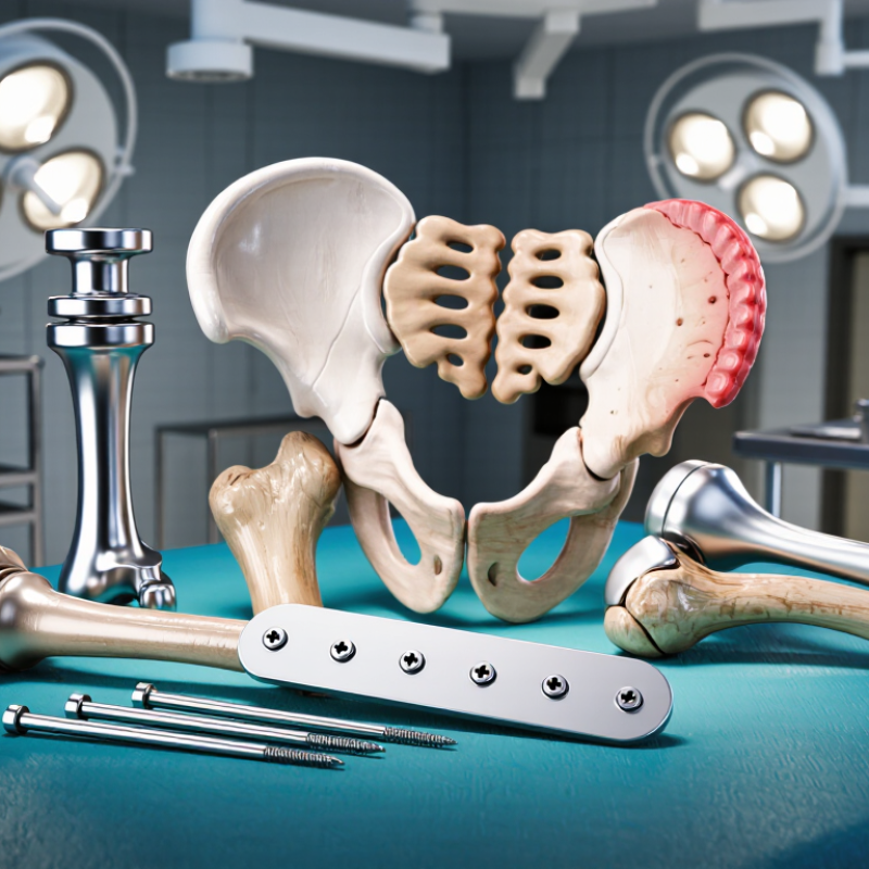
Orthopedic implant breakage means the device has lost structural strength. A plate may crack, a screw may shear, or an intramedullary nail may fatigue through repeated loading. It does not always mean poor surgery. Delayed bone healing, excessive stress, infection, corrosion, and falls can all contribute. Pain, swelling, clicking, or renewed deformity deserve prompt clinical assessment. One detail is easy to miss. Breakage may appear after months of normal movement, not immediately after surgery.
Professional registries provide useful context, but their numbers need careful reading. The UK National Joint Registry’s 21st Annual Report tracks more than four million hip and knee procedures. It uses revision surgery as a major outcome, although revision does not equal implant fracture. The FDA’s MAUDE database collects adverse-event reports, yet it cannot calculate a precise failure rate. Reporting may be incomplete, duplicated, or influenced by legal and clinical factors. The numbers are imperfect. That limitation matters when patients search online.
Tips: Keep scheduled X-rays, especially after fracture fixation. Report increasing pain, warmth, drainage, or a sudden loss of function. Follow weight-bearing instructions closely. Avoid smoking, because nicotine can impair bone healing. Maintain protein, vitamin D, and calcium intake when clinically appropriate. Ask whether bone healing is progressing before returning to running, heavy lifting, or contact activity. Surgeons also need to review alignment, implant loading, infection signs, and nonunion risk at each follow-up. Prevention is shared work, not a single instruction.
Implant breakage usually results from repeated loading, poor bone healing, excessive stress, malposition, infection, or delayed weight-bearing restrictions. The chart shows representative midpoint values from reported clinical ranges; actual rates vary by implant design, anatomy, surgical technique, and follow-up duration.
Prevention focuses on accurate implant positioning, stable fixation, appropriate implant sizing, treatment of infection and osteoporosis, smoking cessation, regular follow-up, and gradual return to activity. Sudden pain, swelling, deformity, or loss of function should be assessed promptly by an orthopedic professional.
Orthopedic implant failure rarely has one cause. Common types include aseptic loosening, infection, wear, dislocation, periprosthetic fracture, and implant breakage. Loosening may begin quietly, as bone weakens around the implant and pain appears during walking. Wear particles can trigger inflammation and osteolysis. Infection may develop early or years later. A sudden fall, poor bone quality, or repeated high-impact loading can fracture an implant or the surrounding bone.
Registry data show why failure rates must be interpreted carefully. The 2024 Australian Orthopaedic Association National Joint Replacement Registry identifies loosening, infection, fracture, and instability among major reasons for revision surgery. The UK National Joint Registry’s 21st Annual Report also shows that ten-year revision risk often falls within roughly 3–5% for many primary hip and knee replacement groups, depending on age, fixation method, implant design, and patient health. These figures are not guarantees. They describe populations, not one person.
Prevention starts with accurate sizing, stable fixation, sterile surgical technique, and realistic activity planning. Surgeons also assess bone density, smoking history, diabetes, and previous infection. Patients should report increasing pain, warmth, drainage, clicking, or sudden instability early. Regular follow-up can detect migration or bone loss before major damage occurs. Still, no checklist prevents every failure. Registry reports cannot fully measure poor adherence, unreported symptoms, or the daily mechanical stresses placed on an implant. The uncomfortable lesson is simple: “successful” surgery still requires long-term monitoring.
Orthopedic implants break when repeated stress exceeds their fatigue strength. A plate may look solid, yet thousands of walking cycles can create a tiny crack. Hip stems, screws, and spinal rods face similar risks. Poor alignment, undersized components, excessive body weight, falls, and delayed bone healing can increase loading. Corrosion or fretting may weaken metal junctions over time. Sometimes, the bone fails first.
Registry evidence shows that mechanical problems are clinically important. The Australian Orthopaedic Association National Joint Replacement Registry 2024 Annual Report reviewed more than 1.8 million joint-replacement procedures. It reported that revision risk differs by implant design, fixation method, patient age, and diagnosis. However, registries usually record revision surgery, not every implant crack. That limitation matters. A “low revision rate” does not mean zero breakage.
Prevention starts before surgery. Surgeons assess bone quality, anatomy, activity level, and expected loads. Accurate sizing and stable fixation reduce concentrated stress around holes, joints, and interfaces. During recovery, gradual weight-bearing gives bone time to unite. Follow-up imaging can reveal loosening, bending, or a changing fracture line before sudden failure. Patients should report new pain, clicking, swelling, or reduced function, especially after a fall. Engineers also use fatigue testing, corrosion analysis, and manufacturing controls, but testing cannot reproduce every human movement. Real-world surveillance remains necessary. Even careful treatment can fail, and that uncomfortable fact should guide better implant design and patient monitoring.
When an orthopedic implant breaks, diagnosis usually begins with a careful timeline. Patients may report new pain, swelling, grinding, reduced movement, or a sudden loss of support. Sometimes, the change is subtle. Pain after a minor twist does not prove breakage, but it deserves assessment. A clinician asks about the original operation, recent falls, activity changes, and earlier symptoms. That history can reveal whether the problem is a broken plate, a loose screw, bone nonunion, infection, or another cause.
During examination, the clinician checks alignment, tenderness, warmth, skin changes, limb length, and nerve or blood-vessel function. Visible deformity or a sharp metal edge under the skin needs prompt medical attention. Standard X-rays are often the first imaging test. Images from several angles can show a fractured component, migration, bending, or poor bone healing. Comparing new films with earlier images is especially useful. However, an implant can appear intact on one view. No single scan tells the whole story. Computed tomography may clarify complex fractures or hidden hardware damage. Magnetic resonance imaging requires careful safety review around metal.
Blood tests may support the investigation when infection is possible. Markers such as C-reactive protein and erythrocyte sedimentation rate can add evidence, but neither test proves the diagnosis alone. Fluid or tissue sampling may be needed before treatment decisions. Diagnosis is not always neat. Pain can come from the implant, the bone, nearby tissue, or all three. Clinical judgment matters. An orthopedic specialist combines symptoms, examination, imaging, and surgical history before recommending revision or observation.
Orthopedic implants may break when repeated stress exceeds their design limits. Poor bone quality, infection, falls, and delayed healing can increase this risk. Excessive weight-bearing too soon may also overload the implant. In some cases, a small crack begins around a screw or joint component.
The patient may notice increasing pain, swelling, clicking, or reduced movement. These symptoms need timely medical assessment.
A careful surgical plan is essential. Surgeons assess bone density, alignment, joint stability, and the patient’s daily demands before choosing an implant. Sterile technique and appropriate fixation help reduce infection and loosening. Patients should follow weight-bearing instructions, even when the limb feels better. Healing often takes longer than expected.
Rehabilitation should be gradual and supervised. Physical therapy can improve strength, balance, and movement control without placing sudden stress on healing tissue.
Avoid smoking, because nicotine can restrict blood flow and delay bone repair. Adequate protein, calcium, vitamin D, and regular medical reviews may support recovery.
Use handrails after surgery, and remove loose rugs at home. A follow-up X-ray can reveal alignment changes before symptoms become severe.
No plan is perfect. I have seen recovery change after one careless step. Report persistent pain, warmth, drainage, or a new deformity promptly.
Do not adjust activity levels without guidance from your orthopedic team. Regular follow-up remains important, even when daily movement seems normal.
Common problems include loosening, infection, wear, dislocation, fracture, and implant breakage. Some develop quietly. Pain during walking may appear before major damage.
Repeated stress can exceed the implant’s design limits. Falls, weak bone, infection, delayed healing, and early heavy loading increase the risk. A small crack may begin near a screw or joint component.
Report increasing pain, swelling, warmth, drainage, clicking, reduced movement, or sudden instability. A new deformity needs prompt medical assessment. Do not simply wait for symptoms to disappear.
Surgeons assess bone quality, alignment, joint stability, and daily physical demands. Accurate sizing and stable fixation are important. Sterile surgical technique can reduce infection risk.
Follow weight-bearing instructions, even when the limb feels better. Rehabilitation should be gradual and supervised. Healing often takes longer than expected.
Avoid smoking because nicotine may restrict blood flow and delay bone repair. Adequate protein, calcium, and vitamin D may support recovery. Handrails and clear floors can reduce fall risks at home.
Regular reviews can identify migration, alignment changes, or bone loss. Follow-up X-rays may show problems before severe symptoms develop. Normal daily movement does not prove everything is stable.
Some registry groups show roughly 3–5% revision risk at ten years. Rates vary with age, fixation, implant design, and health. These figures describe groups, not one person.
No checklist is perfect. A careless step, unexpected fall, or delayed healing can change recovery. The uncomfortable part is simple: successful surgery still requires long-term monitoring.
Can orthopedic implants break? Yes, although modern orthopedic implants are designed to be strong and durable, they can sometimes fracture, loosen, bend, or wear out. Implant breakage may result from repeated stress, poor bone healing, incorrect placement, excessive physical demands, infection, or natural changes in the surrounding bone and tissue. Common failures include broken screws, plates, rods, joint components, and fixation devices. Symptoms may include increasing pain, swelling, instability, reduced movement, unusual sounds, or a sudden loss of function.
Doctors diagnose suspected implant failure through a physical examination, medical history, and imaging tests such as X-rays or other scans. Treatment depends on the type and severity of the problem and may involve observation, activity changes, medication, or revision surgery. Preventive measures include selecting the appropriate implant, ensuring accurate surgical placement, following rehabilitation instructions, maintaining healthy bone strength, avoiding excessive impact during recovery, attending follow-up appointments, and promptly reporting new or worsening symptoms. Proper care and regular monitoring can reduce the risk of implant failure and support long-term recovery.