In the realm of orthopedic medicine, understanding how do doctors check orthopedic implants is crucial. Dr. Emily Tran, an orthopedic surgeon at the renowned Healing Horizons Clinic, states, “Regular monitoring of implants is vital to ensure patient safety and long-term success.” Her expertise highlights the importance of thorough evaluations in detecting any potential issues.
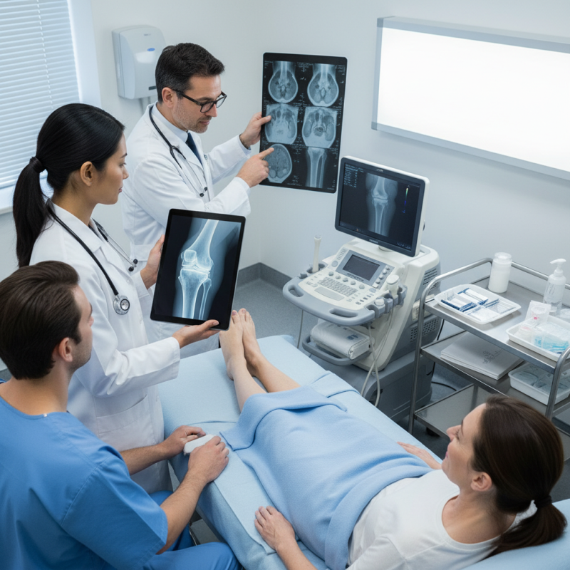
Orthopedic implants, such as joint replacements, require careful assessment. Doctors utilize various methods, including X-rays and MRIs, to check the integrity and position of these devices. They look for signs of wear, displacement, or infection. Regular follow-ups are essential for early detection, as problems can develop slowly over time.
Patients may experience anxiety about the condition of their implants. Many wonder how often these examinations should occur. While the answer can vary, staying proactive is key. Engaging in open communication with healthcare providers can yield clear guidance. Ultimately, understanding how do doctors check orthopedic implants fosters trust in medical care and enhances patient outcomes.
Understanding Orthopedic Implants and Their Importance
Orthopedic implants play a vital role in improving patients' quality of life. These devices, such as plates, screws, and joints, help in stabilizing broken bones and correcting deformities. Their importance cannot be understated, as they often serve as a foundation for recovery in orthopedic procedures.
Doctors utilize various methods to evaluate the integrity of these implants. X-rays are commonly used to check alignment and position. They provide a clear image of how implants interact with the bone. Additionally, CT scans can reveal issues that may not be visible on X-rays. These more detailed scans allow for a thorough assessment of both the implant and surrounding tissues.
Monitoring a patient's progress is essential. Sometimes, patients may experience discomfort or complications. Regular follow-ups ensure that any potential issues are addressed promptly. While these examinations are effective, the outcomes are not always perfect. Each patient's response varies, and unexpected challenges can arise. Continuous evaluation is critical in orthopedic care aimed at enhancing recovery and ensuring long-term success.
Common Techniques for Imaging Orthopedic Implants
Doctors rely on various imaging techniques to assess orthopedic implants after surgery. X-rays are commonly used for initial evaluations. They provide quick visuals, showing the implant's position relative to the bone. However, X-rays only show bone and may miss soft tissue problems.
MRI is another essential tool. It excels in visualizing soft tissues surrounding the implant. Doctors use MRI to check for possible infections or issues with ligaments. Still, MRI can be limited by the presence of metal implants, which may cause artifacts in the images.
CT scans offer detailed cross-sectional views. They help in examining complex fractures or assessing the bone density around the implant. However, CT scans involve higher radiation exposure compared to standard X-rays. Each method has its pros and cons. Adjusting imaging strategies based on specific patient needs can lead to better outcomes.
Considering the limitations of imaging methods is crucial for accurate diagnosis. Findings might sometimes lead to unexpected conclusions. Reflecting on these techniques allows for continuous improvement in patient care.
Top 5 Imaging Techniques for Orthopedic Implants
Evaluating Implant Stability Through Clinical Evaluation
Evaluating implant stability is crucial for the success of orthopedic procedures. A doctor's clinical evaluation typically begins with a thorough patient history. Understanding any pain, mobility issues, or functional limitations is essential. Observations during physical examination play a significant role. Doctors look for signs of swelling, tenderness, or abnormal movement at the implant site.
Imaging techniques are also integral to assessment. X-rays reveal the alignment and integrity of the implant. However, they may not show soft tissue conditions adequately. Advanced methods, such as MRI or CT scans, provide more detailed insights. These techniques can help identify the surrounding bone’s quality and potential complications.
Despite these methods, discrepancies can arise. Some patients may report pain even with well-placed implants. This suggests a need for further investigation into individual factors influencing implant success. Hence, ongoing follow-ups are vital for monitoring potential changes over time. Balancing clinical findings with patient feedback remains a challenge. Continuous learning from these evaluations is a hallmark of effective orthopedic care.
Utilizing Advanced Technologies for Implant Assessment
Orthopedic implant assessment has evolved significantly with the advent of advanced technologies. These innovations allow for more precise evaluation of implant performance and longevity. One prominent method is imaging techniques, including MRI and CT scans. These provide detailed images of bone integration and alignment, which are crucial for identifying potential issues early.
Moreover, biomechanical analysis is becoming vital. This technique assesses how implants respond under real-life conditions. According to a recent study from the Journal of Orthopedic Research, more than 30% of implants show signs of wear within ten years. This statistic emphasizes the need for ongoing evaluation using sophisticated tools.
Another exciting development is the integration of artificial intelligence. AI can analyze vast datasets, predicting failures based on patterns found in previous cases. Yet, this technology isn't foolproof. Data can sometimes lead to misinterpretations, given the variability in individual patient responses. This area requires continued research and refinement to ensure accuracy and reliability in assessment.
Top 5 Ways Doctors Check Orthopedic Implants
| Method |
Description |
Benefits |
Limitations |
| X-ray Imaging |
A widely used technique that provides images of bone and implant positioning. |
Non-invasive and quick, great for initial assessment. |
May not show soft tissue conditions; limited detail on implant integrity. |
| MRI |
Magnetic resonance imaging offers detailed images of both soft and hard tissues. |
Excellent for assessing surrounding soft tissues and detecting inflammation. |
Can be limited by the presence of metal implants which may affect image quality. |
| CT Scans |
Computed tomography provides cross-sectional images for a detailed assessment. |
High-resolution images that can detect minute structural changes. |
Higher radiation exposure compared to X-rays. |
| Ultrasound |
Uses sound waves to produce live images of tissues and structures. |
Safe and real-time imaging; can assess soft tissues around the implant. |
Operator-dependent and may be less effective for deeper structures. |
| Bone Scintigraphy |
A nuclear imaging technique that evaluates bone metabolism and activity. |
Useful for detecting stress fractures and infections early on. |
Involves radiation exposure and may detect unrelated bone activities. |
Monitoring Long-Term Performance of Orthopedic Implants
Monitoring the long-term performance of orthopedic implants is crucial for successful patient outcomes. Studies indicate that nearly 20% of patients experience complications related to implants within the first ten years. Monitoring methods must therefore adapt to ensure these devices function properly over time.
One common approach is using imaging techniques, such as X-rays or MRIs. These methods help in assessing the positioning and integration of the implant. Research shows that regular imaging can detect misalignments or bone resorption early, reducing the risk of further complications. However, the frequency and type of imaging can vary based on individual patient factors, which complicates standardization.
Another method involves the collection of patient-reported outcomes (PROs). A significant 75% of orthopedic surgeons believe PROs provide valuable insights into the implant's effectiveness in daily life. Gathering this data can reveal issues that imaging might not capture, like pain or mobility changes. Adapting to patient feedback is essential but can be challenging. Balancing clinical assessments with patient experiences requires ongoing dialogue between doctors and patients. This dual approach is necessary for advancing orthopedic care.
Conclusion
Orthopedic implants play a crucial role in improving patients' mobility and quality of life after surgery. Understanding how to assess these implants is vital for ensuring their effectiveness and longevity. So, how do doctors check orthopedic implants? They commonly utilize imaging techniques such as X-rays, MRIs, and CT scans to visualize the position and integration of the implants within the bone structure. Clinical evaluations are also essential, where doctors examine the stability and functionality of the implant during physical assessments.
In addition, advanced technologies, such as robotics and digital monitoring systems, are increasingly being implemented for more precise evaluation of implant performance. Long-term monitoring strategies help in assessing the durability of orthopedic implants, allowing for timely interventions when necessary. Ultimately, a combination of these methods enables doctors to effectively check orthopedic implants and ensure optimal outcomes for their patients.